

Science & Tech · Science & Environment · 11 hrs ago
New analysis of Rosalind Franklin’s notes supports her role in DNA research
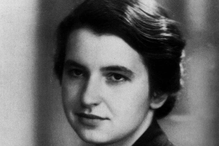
Rosalind Franklin was a British scientist who used X-ray images to study DNA.
Her image 51, made with graduate student Raymond Gosling at King’s College, was seen by James Watson without her permission.
Watson and Francis Crick used information from Franklin’s work while developing a three-dimensional model of DNA in 1953, alongside Maurice Wilkins.
A new study by historian Alistair Sponsel and crystallographer Brian Sutton examines Franklin’s laboratory notes and an earlier X-ray image, numbered 49.
They argue that the notes show she had already considered DNA’s helical shape and that image 51 was made to provide clearer evidence, not because she had missed the structure.
Franklin died in 1958, before Watson, Crick and Wilkins received the 1962 Nobel Prize.
The study adds evidence about Franklin’s contribution, while the story also notes that Watson and Crick made important discoveries of their own.

A new analysis of Rosalind Franklin’s laboratory notebooks argues that she understood DNA’s helical form and made an X-ray image to support her findings.
Historians Alistair Sponsel and crystallographer Brian Sutton published their analysis in the Journal of the History of Biology.
They base their argument on an earlier X-ray image, numbered 49, and Franklin’s notebook entries.
The article says Franklin sought a clearer image for publication, rather than failing to recognize the helix.
Franklin’s research contributed to work by James Watson and Francis Crick, who built a three-dimensional DNA model in 1953.
- Who
- Rosalind Franklin, and the researchers Alistair Sponsel and Brian Sutton.
- What
- An analysis of Franklin’s notes supports her role in recognizing DNA’s helical form and producing X-ray image 51.
- When
- The analysis was reported on October 11, 2026. The article does not state when it was published in the journal.
- Where
- Franklin’s DNA research took place at King’s College. The analysis appeared in the Journal of the History of Biology.
- Why
- Sponsel and Sutton argue that Franklin had already recorded a helical hypothesis and measurements in notes about image 49, and wanted a clearer image for publication.
Sponsel and Sutton
The established interpretation of Franklin’s work
Franklin’s understanding of DNA’s shape
Sponsel and Sutton
Franklin was aware of the helical form and produced image 51 to support her finding.
The established interpretation of Franklin’s work
The established interpretation described in the article says Franklin did not recognize that image 51 showed a helix.
Why image 51 had no notebook entry
Sponsel and Sutton
Franklin wanted a more presentable image, and her earlier notes already contained the relevant hypothesis and measurements.
The established interpretation of Franklin’s work
The lack of notes was interpreted as evidence that she had overlooked the image’s significance.
in each turn of the helix (if there is a helix)
Watson and Crick assembled a three-dimensional model of DNA.
Franklin died.
Watson, Crick and Maurice Wilkins received the Nobel Prize.
Folha reported on Sponsel and Sutton’s analysis of Franklin’s notebooks.
- Researchers
- Alistair Sponsel and Brian Sutton
- Journal
- Journal of the History of Biology
- Earlier image
- X-ray image 49
- Notable image
- DNA X-ray image 51
- Model year
- Watson and Crick assembled their model in 1953
- Nobel Prize year
- 1962
Sources
Related news
Researchers map hair-like sensory cells across squid bodies
Bellbird and Seriema Among the Loudest Birds Measured
Survey finds only 83 mature cheetahs across a vast area of South Africa
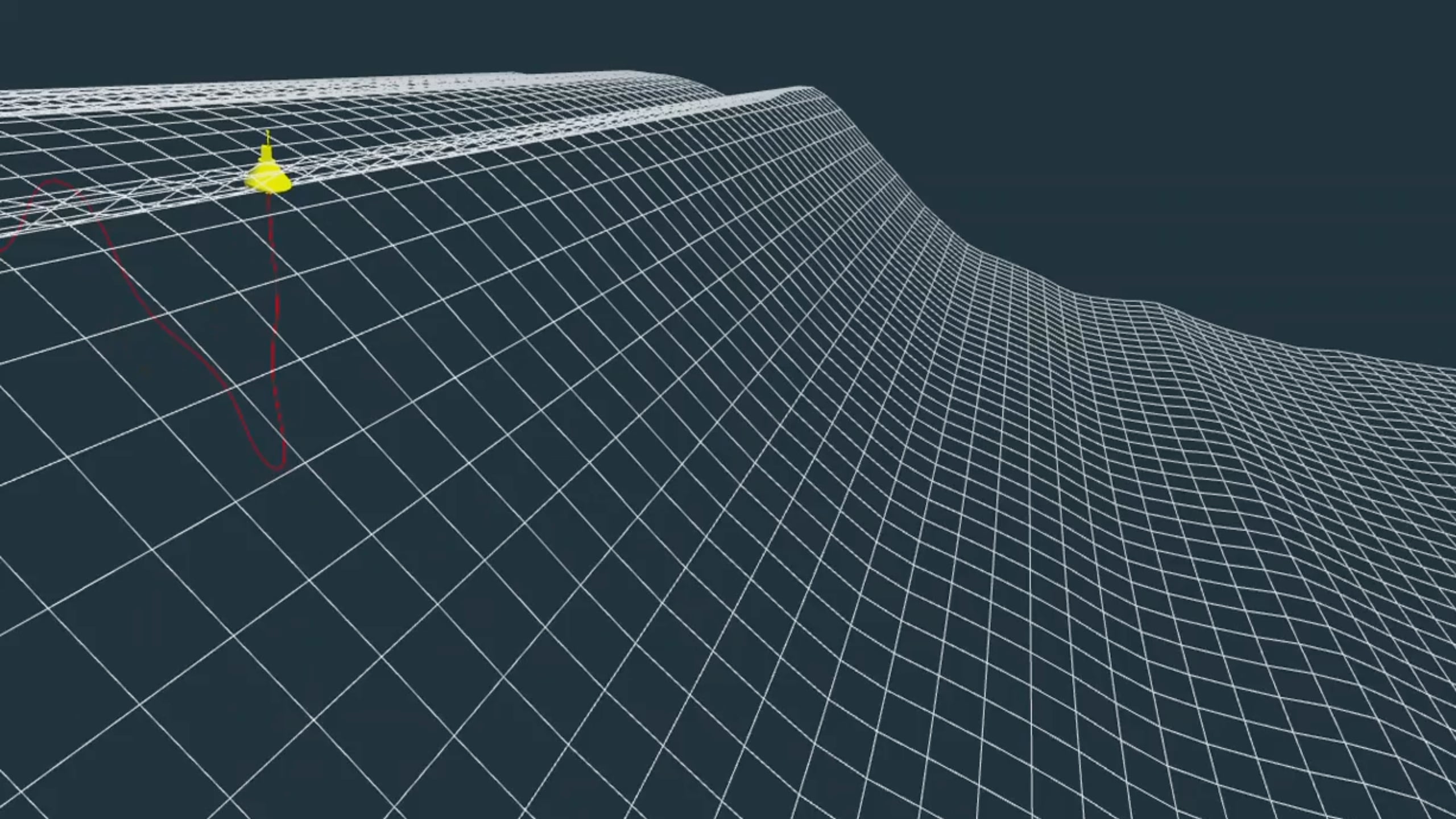
British Columbia rogue wave was nearly three times taller than nearby waves

Detached sea cucumber tissue remained active for more than three years
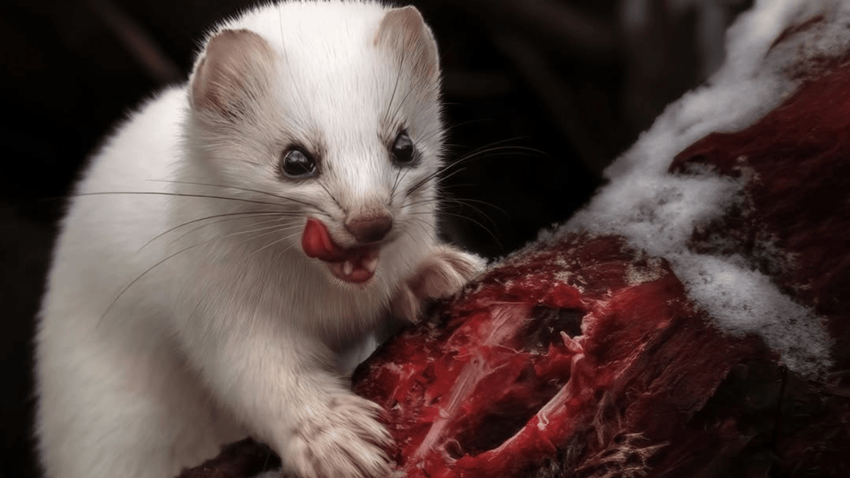
New book examines the survival skills of 75 fierce forms of nature

Researchers confirm a new giant waterlily species in Colombia

Air pollution reaches ‘very poor’ levels in towns near Manaus
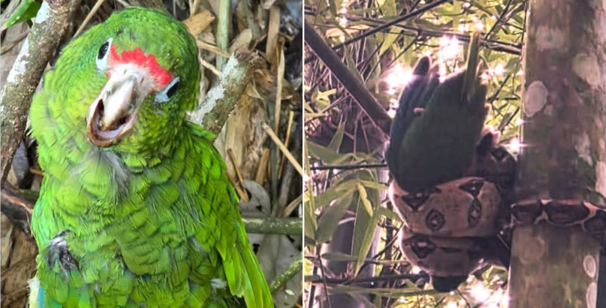
Young Puerto Rican parrot rescued from invasive boa
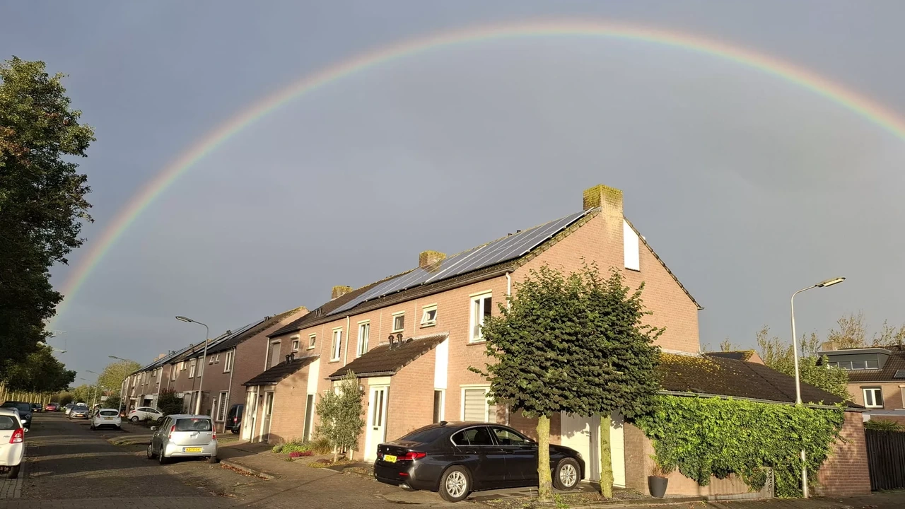
Warm, mostly sunny weather forecast across the Netherlands this week
Showers and morning fog forecast in parts of Türkiye