

Science & Tech · Science & Environment · 3 hrs ago
Scientists capture molecular-scale images of bacterial cell ends
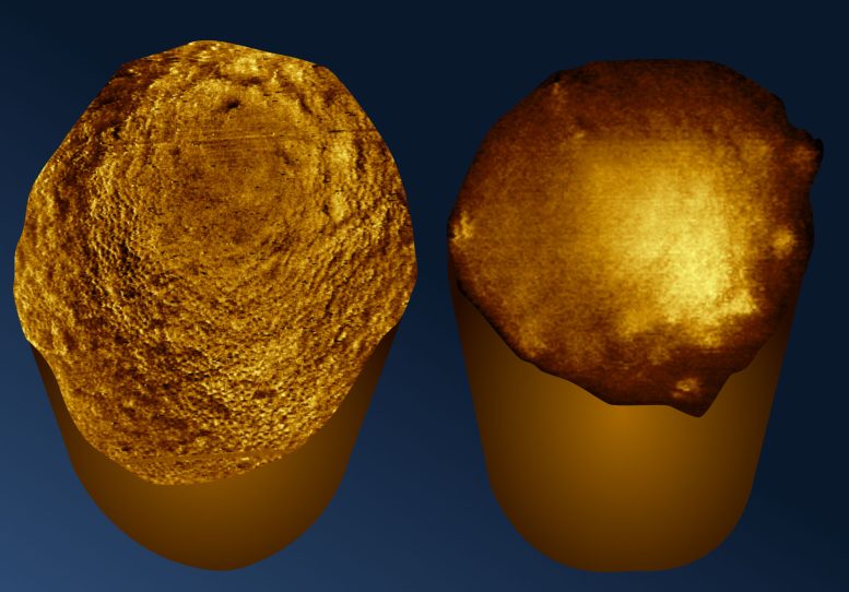
UCL researchers captured molecular-scale images of living bacteria’s ends, called poles, for the first time.
They studied E. coli and two species linked to difficult-to-treat infections, Pseudomonas aeruginosa and Acinetobacter baumannii.
The team held the bacteria against specially treated glass and used atomic force microscopy to map their surfaces.
More active E. coli had folds at its poles, while less active cells generally had smoother, rounded ends.
The other two species lacked tiny surface pores seen on E. coli, showing that bacterial surfaces differ.
These differences could affect how bacteria interact with antibiotics, and some treatments already target cell poles, where bacteria divide.
Researchers still need to establish how these structures affect drug susceptibility, but the method could allow clearer study of living bacteria across more species.

Researchers at University College London captured molecular-resolution images of the ends, or poles, of living rod-shaped bacteria for the first time.
They used atomic force microscopy to examine bacteria held upright on specially treated glass.
The images showed fold-like defects at the poles of rapidly dividing E. coli, while less active cells generally had smoother, rounder poles.
Pseudomonas aeruginosa and Acinetobacter baumannii lacked the network of tiny surface pores observed in E. coli.
The findings may help researchers study bacterial cell division and differences in how species respond to antibiotics, but the effects on drug susceptibility remain uncertain.
- Who
- Researchers at University College London, led by Anna Scheeder and Bart Hoogenboom.
- What
- They captured molecular-resolution images of the poles of living rod-shaped bacteria.
- When
- The study was published on November 18, 2026.
- Where
- University College London; the bacteria were imaged on specially treated glass.
- Why
- To examine bacterial surface structures that could help explain differences between species and their responses to antibiotics.
This story does not have two clearly opposing sides.
Bacteria on the treated glass were generally immobilized horizontally.
Many bacteria had unexpectedly stood upright, exposing their poles for imaging.
The study was published in Nature Communications.
- Researchers
- University College London
- Imaging method
- Atomic force microscopy
- Bacteria examined
- E. coli, Pseudomonas aeruginosa and Acinetobacter baumannii
- Publication
- Nature Communications, November 18, 2026
- Immobilization surface
- Specially treated glass
What people are saying
Our immobilization method opens up exciting new possibilities. We can now study the armor of bacteria across different species more reliably and clearly observe nanoscale differences that were previously difficult to assess.
Imaging the poles of bacteria is like opening a view of the back of the moon.
The poles are where cells divide and so contain critical information about this process.