

Science & Tech · Science & Environment · 3 hrs ago
SPIFFI captures sharper images of moving structures in living cells
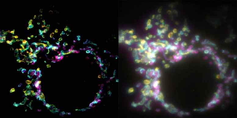
A team at EPFL developed a new imaging method called SPIFFI to study living cells.
Many super-resolution techniques combine hundreds or thousands of images, but moving cell structures can shift during that process and blur the result.
SPIFFI uses four polarization-sensitive images from one exposure to reveal details as small as 160 to 170 nanometers, with up to twice the resolution.
The team used it to image moving mitochondria and microtubules, as well as structures joining and splitting.
Because each exposure produces a sharp image, researchers can make super-resolution videos of live cells instead of assembling a single image from many frames.
The team is now working to make the system more compact and integrate it with three-dimensional microscopy.

Researchers at EPFL have developed SPIFFI, a microscope technique that captures fine details in living cells in a single camera exposure.
The method can improve image resolution by up to twofold and distinguish structures as small as 160 to 170 nanometers.
SPIFFI separates fluorescent light into four polarization-sensitive channels to recover structural detail.
The researchers used it to image moving mitochondria and microtubules, as well as structures undergoing fusion and splitting.
The team is working to make the system more compact and integrate it with three-dimensional microscopy.
- Who
- A team led by Aleksandra Radenovic at EPFL’s Laboratory of Nanoscale Biology developed SPIFFI.
- What
- The technique captures super-resolved images of cellular structures in a single camera exposure.
- When
- The research was published in Nature Methods; the article was published on 2026-10-30.
- Where
- At the Laboratory of Nanoscale Biology in EPFL’s School of Engineering.
- Why
- To capture moving structures in living cells without the blurring that can result from methods requiring many images.
This story does not have two clearly opposing sides.
SciTechDaily published its report on the SPIFFI research.
The team is working to make the system more compact and integrate it with three-dimensional microscopy.
- Technique
- Spatial Polarization-Induced Fluorescence Fluctuation Imaging (SPIFFI)
- Resolution improvement
- Up to twofold
- Smallest structures distinguished
- 160 to 170 nanometers
- Imaging channels
- Four polarization-sensitive channels
- Research institution
- Laboratory of Nanoscale Biology, EPFL
What people are saying
Essentially, previous approaches used temporal information to resolve spatial resolution, but this doesn’t work very well on living cells
With previous techniques, taking many images would only result in one super-resolved frame. With SPIFFI, every frame is super-resolved, meaning we can now produce super-resolution videos of live cells
Our experiments show that SPIFFI can capture fast-moving processes within cells, while enabling high-throughput, multi-dimensional imaging beyond the limits of conventional microscopes